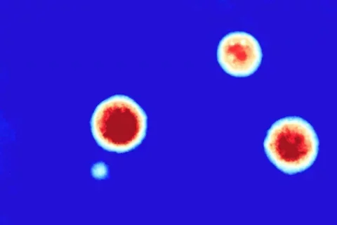
UCLA Researchers Develop New Way to Grow and Measure How Tumor Organoids Respond to Therapy

Scientists from the Teitell and Soragni labs, both part of the UCLA Jonsson Comprehensive Cancer Center, have developed a novel method for bioprinting miniature tumor organoids that closely mimic the architecture and function of real tumors. This advancement allows for detailed analysis of individual organoids using advanced imaging techniques. The team utilized a bioprinting technique, combining it with high-speed live cell interferometry and machine learning algorithms to measure and analyze thousands of organoids simultaneously. This non-destructive method enables the identification of organoids' sensitivity or resistance to specific therapies, facilitating the selection of effective treatment options for patients. Dr. Michael Teitell, the director of the UCLA Jonsson Comprehensive Cancer Center and co-senior author of the study (along with Dr. Alice Soragni), emphasized the importance of this approach in identifying personalized treatments for individuals with rare or difficult-to-treat cancers.
The researchers plan to apply this approach to organoids from hard-to-treat rare cancers, aiming to uncover novel therapeutic avenues and resistance mechanisms for personalized treatment strategies. The study was supported by grants from various sources, including the National Institutes of Health and the Department of Defense, and published in the journal of Nature Communications. Co-first authors include Peyton Tebon, Bowen Wang (graduate student in the Teitell lab), and Alexander Markowitz, with contributions from other UCLA researchers.